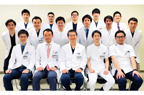


The Kobe University Orthopaedic Sports Medicine and Lower Extremity Division is served by five faculties: Prof. Ryosuke Kuroda, Dr. Takehiko Matsushita, Dr. Yuichi Hoshino, Dr. Noriyuki Kanzaki, and Dr. Daisuke Araki. Our treatment strategy is to restore normal joint function and kinematics by using comprehensive joint preserving techniques, such as ligament reconstruction, osteotomy, meniscal repair, or cartilage repair, including cell therapies. Once injuries occur, our sports medicine experts use the most advanced methods to evaluate and treat injuries in professional, nonprofessional, and recreational athletes of all ages. Our mission is returning patients to an active lifestyle based on our clinical and basic researches.
Anterior cruciate ligament (ACL) and knee ligament reconstruction
Our division traditionally concentrates on the safe return to sports after ACL or knee ligament injuries. Once ACL is injured, ACL reconstruction is the gold standard for treatment due to its poor self-healing ability. To quantitatively assess knee joint laxity, we have developed the three-dimensional (3-D) electromagnetic measurement system (EMS) in 2004 and used this system to patients with knee ligament injury. This system can evaluate the Lachman and pivot shift tests non-invasively and quantitatively. Based on several biomechanical researches, anatomic double-bundle ACL reconstruction, which reproduces both the anteromedial and posterolateral bundles, is our first choice for patients with ACL injury. In addition, we also have some treatment options, such as bone-patellar tendon-bone graft ACL reconstruction with Kurosaka screws, selective single-bundle ACL reconstruction (ACL remnant preserved augmentation technique) or epiphyseal plate-preserving technique. These clinical researches have led many athletes to undergo surgical treatment in our division.
Articular cartilage injuries
For focal osteochondral lesions or osteochondritis dissecans, we select the treatment strategies, such as arthrosopic bone marrow stimulations (drilling, micro-fracture or abrasion chondroplasty), autogenous osteochondral graft transfer (mosaicplasty), or autologous chondrocyte implantation, based on osteochondral lesion size. To date, we have reported good clinical results of these treatments. For autogenous osteochondral graft transfer, we investigated the ideal matching patterns of articular surface profiles of the donor and recipient sites using a 3-D laser scanning method. For larger traumatic cartilage defects, autologous chondrocyte implantation is the first choice. Since 2012, we conducted the investigator-initiated clinical research of the 3rd generation of this treatment in collaboration with the Institute of Biomedical Research and Innovation Hospital (http://chuo.kcho.jp/foreign_page/eng/eng_). The good outcome of this clinical research is now stepped onto the sponsor-initiated clinical research. We recently performed the 2nd generation of autologous chondrocyte implantation using JACCR (J-TEC, Japan), with good post-operative outcomes.
Meniscal injuries
To treat meniscal injuries, we are actively performing arthroscopic meniscal repair because meniscectomy leads to a high risk of osteoarthritis. Furthermore, we used fibrin clots for meniscal repair to enhance the clinical results. In animal studies, we reported that platelet-rich plasma (PRP) enhances the healing of meniscal defects. Based on this result, we will also consider clinical trials or either surgical treatments using PRP or intracapsular injection, with the goal of further improving our results.
Patellofemoral joint disorders
Patellar dislocation is a multifactorial problem because patellar stability relies on limb alignment, the osseous structure of the patella and trochlea, and the integrity of static and dynamic soft-tissue constraints. To treat these patellar dislocations, our first choice is medial patellofemoral ligament (MPFL) reconstruction. This technique is an operative procedure independently developed at our division, which has shown good clinical results. Furthermore, by developing 3-D analysis software in collaboration with the Kobe University Faculty of Engineering, we perform motion analysis of normal knees, adding the individual anatomy of each case to preoperative planning, with the aim of further improving postoperative results.
Early-stage osteoarthritis
As the sports population is growing, the importance of joint preservation surgery is highlighted. Recently, we actively select osteotomy, which improves pain and disability from isolated unicompartmental disease in the knee through surgical correction of weight-bearing alignment, as a treatment option for highly active and/or relatively young patients with osteoarthritis. Osteotomy includes distal femur osteotomy, high tibial osteotomy (open-wedge or hybrid), and double-level osteotomy, which combines the previous two procedures. The osteotomy type is selected by the deformity of each case. Our division has used computer navigation systems for obtaining more accurate alignment and advancing the state of our treatments.


