 I. The origin of WHHL rabbits
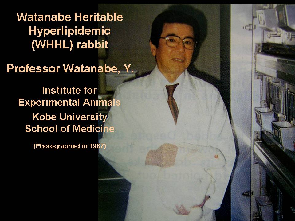
Dr. Yoshio Watanabe (1926-2008), a former professor at our institute, discovered a male Japanese white rabbit in 1973 that showed hyperlipidemia despite feeding on a normal standard diet ( Exp Anim 1977; 26: 35-42 (Japanese); Bull Azabu Vet Coll 1977; 2: 99-124 (Japanese); Atherosclerosis 1980; 36: 261-268). He confirmed the hyperlipidemia of this rabbit was inherited recessively, and started to develop a new rabbit strain showing hyperlipidemia.At that time, few researchers focused on hyperlipidemia in Japan, but late Professor Yoshio Watanabe focused on the importance of development of model animals with hyperlipidemia. We admire the foresight of late Professor Yoshio Watanabe.
For a summary of the history of WHHL rabbit development please refer to the PDF file:Chronology of WHHL rabbits
II. Development of the WHHL rabbit strain
Dr. Watanabe mated this mutant rabbit with 10 normal Japanese white. He repeatedly backcrossed this mutant rabbit and his child rabbits of the rabbits and crossed the child rabbits. He finally established a new rabbit strain with hyperlipidemia in 1979 and named it the WHHL (Watanabe heritable hyperlipidemic) rabbit ( Atherosclerosis 1980; 36: 261-268). After the establishment the WHHL rabbit strain, Dr. Watanabe began to provide WHHL rabbits to many researchers all over the world.
In the WHHL rabbit at the time of the establishment, atherosclerosis occurred in the aorta, but incidence in the coronary lesions was extremely low. Therefore, to increse the incidence of coronary lesions, he performed selective breeding from 1980 to 1984. Consequently, the incidence of coronary lesions was increased.(Atherosclerosis 1985; 56: 71-79).
However, the degree of coronary lesions was mild. Therefore, we perforemd selective breeding again between 1985 and 1991 to increase the degree of the coronary stenosis. After the second selective breeding, WHHL rabbits have suffered from severe coronary atherosclerosis (Atherosclerosis 1992; 96: 43-52). Presently this WHHL rabbit strain is called "coronary atherosclerosis-prone WHHL rabbits".
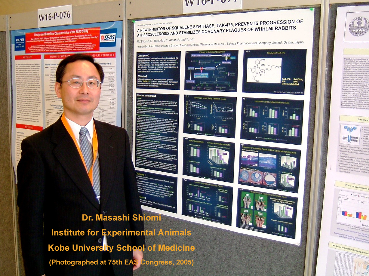 After retirement of Professor Watanabe at the age limit in 1990, Dr. Masashi Shiomi succeded to his WHHL rabbit colony and his studies. Although coronary atherosclerosis-prone WHHL rabbits suffered from severe coronary stenosis, the incidence of spontaneous myocardial infarction was extremely low, and these WHHL rabbits could not be used for studies of myocardial infarction. Since 1994, we have attempted to develop an animal model for spontaneous myocardial infarction by serial and selective breeding of the coronary atherosclerosis-prone WHHL rabbits. After six years of the selective breeding, we developed a new WHHL strain for spontaneous myocardial infarction and designated this myocardial infarction-prone WHHL rabbit strain as the WHHLMI rabbit (Arterioscler Thromb Vasc Biol 2003;23 (7): 1239-1244); Commentary to the International Atherosclerosis Society, IAS Website (http://www.athero.org/comm-index.asp) September 5, 2003.; J Atheroscler Thromb 2004 Sep; 11 (4): 184-189 (See section of "IV. The characteristics of WHHLMI rabbis")
III. Characteristics of WHHL rabbits
1. Lipid metabolism
1). LDL receptor
Similar to human Familial Hypercholesterolemia, LDL receptor function is genetically reduced in WHHL rabbits and they show hypercholesterolemia (FEBS Lett 1980; 118: 81-84; Eur J Biochem 1981; 118: 557-564; Proc Natl Acad Sci USA 1981; 78: 2268-2272; J Biol Chem 1981; 256: 9789-9792).
This defect arises from an in-frame deletion of 12 nucleotides that eliminates four amino acids from the cysteine-rich ligand binding domains of the LDL receptor (Science 1986; 232: 1230-1237). A recent study reported deletion of 11 base pairs (Sci Rep 2016 Jun 1;6:26942).
From this mutant gene, although the precursor of LDL receptor proteins are synthesized normally, the maturation of the LDL receptor protein is delayed and is not transported to the cell surface at a normal rate (Mol Biol Med 1983; 1: 353-367).
2). Plasma lipid level
In our present WHHL rabbits, the average plasma cholesterol levels are about 1,100 mg/dl when they are below 6 months old, about 900 mg/dl at 12 months old, and about 800 mg/dl at 18 months old.
The average plasma triglyceride levels are between 150 and 300 mg/dl.
About 70% of the cholesterol is distributed in LDL fractions, and only a few % in HDL fractions.
3). VLDL secretion from liver
The rate of accumulation of apoB-100 does not differ in perfusates of liver from normal and WHHL rabbits and little or no apoB-48 is accumulated in VLDL fractions (Proc Natl Acad Sci USA 1983; 80: 6096-6100).
ApoB-containing lipoproteins other than VLDL are not secreted from the liver of WHHL rabbits (Proc Natl Acad Sci USA 1983; 80: 6096-6100). An inhibition of the hepatic microsomal triglyceride transfer protein (MTP) activity diminished the VLDL secretion and reduced the plasma LDL levels in homozygous WHHL rabbits. This suggests that MTP inhibitors should have lipid-loweringeffects against homozygous familial hypercholesterolemia. (Eur J Pharmacol 2001; 431: 127-131 ).
4). Activity of enzymes related to lipoprotein metabolism
The plasma CETP activity of WHHL rabbits is about 2 to 3 times higher than that of normal rabbits (Arteriosclerosis 1986; 6:345-351). High CETP activity in WHHL rabbits may be the reason for low HDL cholesterol.
Although the LPL activity of WHHL rabbits is similar to heterozygous WHHL rabbits, the HTGL activity is about 2 times higher than that of the heterozygotes (Biochem J 1990; 272: 647-651). Compared to normal rabbits, in WHHMI rabbits, LPL is about 1.5 times higher and HTGL is about 5 times higher.In rabbits, unlike mice, there is little HTGL activity in plasma before administration of heparin and it is similar to humans.(Exp Anim 2019;68:267-275)
Although the LPL activity of WHHL rabbits is similar to heterozygous WHHL rabbits, the HTGL activity is about 2 times higher than that of the heterozygotes (Biochem J 1990; 272: 647-651). Compared to normal rabbits, in WHHMI rabbits, LPL is about 1.5 times higher and HTGL is about 5 times higher.In rabbits, unlike mice, there is little HTGL activity in plasma before administration of heparin and it is similar to humans.(Exp Anim 2019;68:267-275)
5). Chylomicron metabolism
In a kinetic study of 125I-labeled chylomicrons, the fractional catabolic rate in WHHL rabbits was similar to normal rabbits (Proc Natl Acad Sci USA 1982; 79: 3623-3627).On the other hand, in WHHL rabbits, catabolism of chylomicron is delayed, and there are reports that abnormality of LDL receptor is involved as a cause (J Biol Chem 1995; 270(15): 8578-8587).
6). Low HDL level
Compared to normal rabbits, the fractional catabolic rate of HDL apoA-I in WHHL rabbits is as high as 1.7 times, and the synthetic rate is reported to be reduced to about 50% (Atherosclerosis 1989; 79: 225-230). It may also be related to the fact that CETP activity in blood is as high as 2-3 times that of normal rabbits(Arteriosclerosis 1986; 6:345-351).
7). Age dependent decrease in plasma lipid levels
The plasma cholesterol levels of WHHL rabbits are gradually decreased with age (Metabolism 2000; 49: 552-556).
The HMG-CoA reductase activity of the liver microsomal fraction is increased above 12 month-old. The ACAT activity is decreased between 2 month-old and 6 month-old and remains constant thereafter.
The activity of cholesterol-7a hydroxylase is almost constant.
The cholesterol contents in the whole liver and the microsome fraction are constant.
The VLDL secretion rate is decreased with age.
2. Atherosclerosis
1). Aortic lesion
In current WHHLMI rabbits aortic atherosclerosis is observed from 2 months old despite usual standard rabbit feeding.
The lesions develop first at the orifices of arterial branches.
At 6 month-old, aortic lesions expanded to about 40% of the aortic surface.
At 12 month-old, the lesions cover about 70% of the aortic surface.
Above 18 month-old, the aortic lesions cover most of the aorta.
Regarding lesional composition (Arterioscler Thromb 1994; 14: 931-937; J Atheroscler Thromb 1994; 1: 45-52), in early lesions, many macrophages and few SMCs are observed in the intima and some macrophages penetrate into the arterial media. In transitional lesions observed at about 12 months old, large foam cells derived from macrophages, are increased in the deep area of the intima, and a fibromuscular cap covers the surface. Above 18 months old, cellular components are decreased, but collagen fibers, extracellular lipid accumulation, and cholesterol cleft are increased. In some case, calcium accumulation is also observed in these advanced lesions.
Regarding lesional composition (Arterioscler Thromb 1994; 14: 931-937; J Atheroscler Thromb 1994; 1: 45-52), in early lesions, many macrophages and few SMCs are observed in the intima and some macrophages penetrate into the arterial media. In transitional lesions observed at about 12 months old, large foam cells derived from macrophages, are increased in the deep area of the intima, and a fibromuscular cap covers the surface. Above 18 months old, cellular components are decreased, but collagen fibers, extracellular lipid accumulation, and cholesterol cleft are increased. In some case, calcium accumulation is also observed in these advanced lesions.
2). Coronary lesion
Occurence of coronary lesions
A severe arteriosclerotic lesion was observed in the coronary artery in a mutant rabbit that was the origin of the WHHL rabbit, but as a result of mating with a normal rabbit (JW rabbit) during the development of WHHL rabbit strain, incidence of coronary artery lesions at that time established as a lineage was low rate. After the results of selective breeding(see the section of "Development of the WHHL rabbit strain"), coronary lesions were observed from 2 months old even with feeding of standard chow. The lesions develop mainly at the main stem of the left coronary artery, the origin of the right coronary artery, and the left circumflex arteries. In rabbits, the left circumflex artery is larger and longer than the left anterior descending artery (J Ateroscler Thromb 2018; 25:393-404). In WHHLMI rabbits, the average coronary narrowing (% of plaque area in the area surrounding the internal elastic lamina) of the circumflex artery is about 50% at 6 months old, about 70% at 12 months old, and about 80% above 18 months old(Exp Anim 2004;53:339-346).
Components of coronary lesions
Lesional composition of coronary plaques is relatively fibromuscular compared with the aortic lesions (Arterioscler Thromb 1994; 14: 931-937; Exp Anim 2004;53:339-346). However, several WHHL rabbits show macrophage-rich coronary plaques.
Atherosclerotic coronary arteries exhibited the enhancement in contraction and Ca2+ mobilization in response to serotonin. The 5-HT1B receptor, which is upregulated by atherosclerosis, most likely mediates the augmenting effects of serotonin. (Circulation 2001; 103: 1289-1295)
Outward remodeling of coronary arteries
Atherosclerotic coronary arteries show outward remodeling and coronary atherosclerosis-prone WHHL rabbits are useful animal for studies about coronary outward remodeling. We demonstarted the novel insights into coronary lumen preservation during progression of coronary atherosclerosis (Coronary Artery Disease 2004 Nov; 15(7): 419-426);
Correlation between coronary artery running pattern and severity of coronary lesions
Progression of coronary artery lesion has individual differences. It is suggested that the individual difference is related to the running pattern of coronary arteryiesÅiJ Atheroscler Thromb 2018;25:393-404).
Serum markers for coronary lesions
Serum markers specific for coronary artery lesions were identified in a study using WHHLMI rabbits.(Atherosclerosis 2019;284:18-23).
3). Cerebral artery lesions
4). Other arterial lesions
IV. The characteristics of WHHLMI rabbit
(Arterioscler Thromb Vasc Biol 2003;23 (7): 1239-1244); Commentary to the International Atherosclerosis Society, IAS Website (http://www.athero.org/comm-index.asp) September 5, 2003.; Exp Anim 2004; 53 (4): 339-346; J Atheroscler Thromb 2004 Sep; 11 (4): 184-189)
V. Other property
VI. Inheritance mode
1. Hypercholesterolemia
2. Coronary atherosclerosis
VII. Acute Coronary Syndromes
In humans, it is thought that coronary pulaques are prone to rupture as a part of the fibrous capsule covering the large lipid core is thinned, and these plaques are said to be an unstable plaque leading to sudden cardiac death. In WHHLMI rabbits, such unstable lesions were observed in the coronary arteries, but ruptured coronary plaques could not be found.Therefore, we thought that secondary factors are necessary to cause unstable coronary lesions to rupture, and attempted to induce rupture of coronary lesions by evoking coronary spasm (Arterioscler Thromb Vasc Biol. 2013 Nov; 33(11): 2518-2523).
When bolus intravenous injection of ergonovine under continuous infusion of norepinephrine, ST elevation and ST decline are observed in the electrocardiogram, and these changes are normalized by administration of nitroglycerin. Reduction and recovery of coronary blood flow can be confirmed by coronary angiography. These changes inidicate the occurrence of coronary spasm. In echocardiography, the systolic ventricular diameter increases when spasm occurs, causing heart contraction failure. Serum myocardial ischemic markers (H-FABP, cTroponin-I, Myoglobin) increase 4 hours after the occurrence of spasm.
In coronary lesions, the eruption of macrophages from the clefts of the endothelial cells into the lumen was observed, and the formation of thrombus accompanied by the rupture of the coronary lesion was observed although it was a small number.
VIII. Other abnormal findings
IX. Species difference (Comparison with transgenic or KO mice)
Please see the table summarizing species differences: Essence of species difference about lipoprotein metabolism, atherosclerosis, and myocardial characteristicsÅiPDFÅj
Lipoprotein metabolism and arterial lesions in rabbits are similar to thaose of a humans, but mice differ significantly from humans. Characteristics of rats are similar to mice.
1) Lipid metabolism
2) Atherosclerosis
3) Myocardial characteristics
X. Studies using WHHL rabbits or WHHLMI rabbits
A severe arteriosclerotic lesion was observed in the coronary artery in a mutant rabbit that was the origin of the WHHL rabbit, but as a result of mating with a normal rabbit (JW rabbit) during the development of WHHL rabbit strain, incidence of coronary artery lesions at that time established as a lineage was low rate. After the results of selective breeding(see the section of "Development of the WHHL rabbit strain"), coronary lesions were observed from 2 months old even with feeding of standard chow. The lesions develop mainly at the main stem of the left coronary artery, the origin of the right coronary artery, and the left circumflex arteries. In rabbits, the left circumflex artery is larger and longer than the left anterior descending artery (J Ateroscler Thromb 2018; 25:393-404). In WHHLMI rabbits, the average coronary narrowing (% of plaque area in the area surrounding the internal elastic lamina) of the circumflex artery is about 50% at 6 months old, about 70% at 12 months old, and about 80% above 18 months old(Exp Anim 2004;53:339-346).
Components of coronary lesions
Lesional composition of coronary plaques is relatively fibromuscular compared with the aortic lesions (Arterioscler Thromb 1994; 14: 931-937; Exp Anim 2004;53:339-346). However, several WHHL rabbits show macrophage-rich coronary plaques.
Atherosclerotic coronary arteries exhibited the enhancement in contraction and Ca2+ mobilization in response to serotonin. The 5-HT1B receptor, which is upregulated by atherosclerosis, most likely mediates the augmenting effects of serotonin. (Circulation 2001; 103: 1289-1295)
Outward remodeling of coronary arteries
Atherosclerotic coronary arteries show outward remodeling and coronary atherosclerosis-prone WHHL rabbits are useful animal for studies about coronary outward remodeling. We demonstarted the novel insights into coronary lumen preservation during progression of coronary atherosclerosis (Coronary Artery Disease 2004 Nov; 15(7): 419-426);
| ÅE | Prior quantitative analysis of coronary artery compensatory remodeling is limited by individual variation of arterial size and arterial tapering. Therefore, we developed a new analytical method resolving this limitation and analyzed coronary outward remodeling using perfusion-fixed coronary arteries of WHHL rabbits, an animal model for spontaneous coronary atherosclerosis. In the new analysis, we evaluated how lumen area or arterial size was changed with accumulating atherosclerotic plaques compared to before plaque developed. The lumen area modestly decreased below 10% cross-sectional narrowing (CSN), remained constant from 10% to 68% CSN, and diminished sharply despite continued remodeling above 70% CSN. Up to 70% CSN, arterial remodeling progressed quantitatively to maintain constant arterial wall shear stress as well as lumen area. Quantitative analysis eliminating the previous limitation provides the novel insight that coronary outward remodeling in atherosclerosis maintains lumen size up to 70% CSN in proportion to wall shear stress. |
| ÅE | There is a phenomenon that it can be inferred that the arterial media become thin at the site where macrophages accumulate in the deep layer of the intima lesion and that the fibroblastoid cells change to smooth muscle cells in the arterial adventitia(Atherosclerosis 2008;198:287-293).From this observation, it seems that the thin artery media may reinforce from the adventitial side of the artery. |
Correlation between coronary artery running pattern and severity of coronary lesions
Progression of coronary artery lesion has individual differences. It is suggested that the individual difference is related to the running pattern of coronary arteryiesÅiJ Atheroscler Thromb 2018;25:393-404).
Serum markers for coronary lesions
Serum markers specific for coronary artery lesions were identified in a study using WHHLMI rabbits.(Atherosclerosis 2019;284:18-23).
| ÅE | At 4 months old, which corresponded to when coronary lesions were generated, serum lysophosphatidylcholine (LPC) 22:4 levels were high and diaclyglycerol 18:0-18:0 were low in WHHLMI rabbits with severe coronary lesions. |
| ÅE | At 8months old, which corresponded to when coronary lesions rapidly progressed, citric acid plus isocitric acid, pyroglutamic acid, LPC 20:4 (sn-2), and Cer d18:1-18:2 were high in WHHLMI rabbits with severe coronary lesions. |
| ÅE | At 16 months old, which corresponds to when the coronary artereis were slmost ocluded and the development of myocardial ischemia, serum Phosphatidylethanolamine plasmalogen 16:1p-22:2 was high in WHHLMI rabbits. |
3). Cerebral artery lesions
Although cerebral atherosclerosis was not observed in WHHL rabbits before 1995, WHHL rabbits prone to coronary atherosclerosis (see the section of "Development of WHHL rabbit strain") suffer cerebral atherosclerosis spontaneously in nine months or more without inducing high blood pressure(Atherosclerosis 2001; 156 (1): 57-66 ).
These lesions are mainly observed in the vertebral arteries, the basilar artery, the confluence of vertebral arteries, and the bifurcation of the basilar artery.
However, no lesions were observed in the penetrating arteries.
The severity of the lesion is relatively mild and the lesion is fibromuscular.
The development of cerebral atherosclerosis is not associated with blood pressure in our WHHL rabbits.
4). Other arterial lesions
Atherosclerotic lesions are also observed in the pulmonary artery, carotid arteries, renal arteries, mesenteric artery, celiac artery, and other arteries, while atheromatous lesions are not observed in any small arteriesÅiExp Anim 2001;50:423-426; Exp Anim 2019;68:293-300Åj.
The composition of carotid artery lesion is similar to that of coronary artery lesion, and various lesions (atherosclerosis, fibrotic lesion, stratified lesion etc.) occur in the branch from 6 months of age.
A macrophage-rich lesion develops in the pulmonary artery, and fibrotic lesions are found in the renal artery and the iliac artery-femoral artery. Atherosclerotic arteries and superior mesenteric arteries develop atherosclerosis.
In addition, when the age is over 20 months old, macrophage-derived foam cells are found in the aortic valve, thickening of the aortic valve progresses due to aging, calcium accumulation is also observed, the aortic area decreases, and aortic valve stenosis develops (Atherosclerosis 2018;273:8-14).
IV. The characteristics of WHHLMI rabbit
(Arterioscler Thromb Vasc Biol 2003;23 (7): 1239-1244); Commentary to the International Atherosclerosis Society, IAS Website (http://www.athero.org/comm-index.asp) September 5, 2003.; Exp Anim 2004; 53 (4): 339-346; J Atheroscler Thromb 2004 Sep; 11 (4): 184-189)
| ÅE | The WHHLMI rabbit is the first rabbit model for spontaneous myocardial infarction with restricted feeding of standard chow (Arterioscler Thromb Vasc Biol 2003;23 (7): 1239-1244))ÅD |
| ÅE | WHHLMI rabbits started to die suddenly from 11 months old without apparent symptoms. Histological analysis revealed that myocardial infarction was observed in 97% of rabbits that died up to 35 months old (Exp Anim 2004; 53 (4): 339-346). |
| ÅE | The myocardial lesions were widely distributed from the left ventricle, right ventricle, and ventricular septum where coronary arteries showed severe coronary stenosis (Exp Anim 2004; 53 (4): 339-346). |
| ÅE | Based on the anatomical location, myocardial infarction is classified into subendocardial infarction, intramural infarction, transmural infarction, and subepicardial infarction (Arterioscler Thromb Vasc Biol 2003;23 (7): 1239-1244)). |
| ÅE | In many WHHLMI rabbits, old myocardial infarction was accompanied by fresh myocardial lesions (Arterioscler Thromb Vasc Biol 2003;23 (7): 1239-1244)). |
| ÅE | In WHHLMI rabbits above 20 months of age, 73% of LCX segments show cross-sectional narrworking greater than 90%. |
| ÅE | The electrocardiograms from a WHHLMI rabbit monitored immediately before sudden decease showed the typical change of acute myocardial infarction in humans (Arterioscler Thromb Vasc Biol 2003;23 (7): 1239-1244)). |
| ÅE | Å@These results suggest that in WHHLMI rabbits myocardial ischemia occurs repeatedly as the coronary lesion progresses, and suddenly dies at the last myocardial ischemic event. |
| ÅE | Compared to WHHL rabbits before selection breeding, both aortic lesions and coronary artery lesions are progressed remarkably (Exp Anim 2004; 53 (4): 339-346)ÅB |
| ÅE | Serum total cholesterol increased by about 200 mg / dl compared to WHHL rabbits before selectibe breeding, but the triglyceride levels are comparable. |
| ÅE | Serum total cholesterol level is higher in females than males, but there is no gender difference between aortic lesion, coronary artery lesion, and myocardial infarction(Exp Anim 2004; 53 (4): 339-346)ÅB |
| ÅE | Serum total cholesterol did not correlate with the occurrence of myocardial infarction, but coronary cross-sectional narrwowing correlates with the occurrence of myocardial infarction (Exp Anim 2004; 53 (4): 339-346). |
| ÅE | Culprit coronary arteries have obstructive lesions with large lipid cores covered with a thin fibrous cap, calcium deposits and bleeding within lesions (Arterioscler Thromb Vasc Biol 2003;23 (7): 1239-1244)). |
| ÅE | Å@Despite such unstable plaques, there is no plaque rupture or mural thrombus observed in human acute coronary syndrome. This suggests that the involvement of secondary factors is necessary for rupture of unstable plaques.(Exp Anim 2017; 66 (2): 145-157) |
| ÅE | Various lesions are found in the coronary arteries; atherosclerotic lesions, unstable lesions consisting of a large lipid core covered by a thin fibrous cap, fibrous lesions, lesions with fibrous layers and layers of macrophages and lipid components, fatty plaques, lesions with calcium deposits and vasa vasorum (Exp Anim 2017; 66 (2): 145-157 |
| ÅE | WHHLMI rabbits will contribute to studies of regeneration of cardiac muscle by gene therapy. |
| ÅE | WHHLMI rabbits are useful for studying the mechanism of plaque stability and plaque rupture, or acute coronary syndrome will occur in WHHLMI rabbits by stimulating with other risk factors. |
| ÅE | The aortic and coronary plaques progressed markedly compared to the WHHL rabbits before selective breeding (Exp Anim 2004; 53 (4): 339-346). |
| ÅE | Å@Despite no difference in aortic lesion area and serum lipid level, the age at which myocardial infarction develops and the progression of coronary artery lesion are greatly different, suggesting that there are coronary - specific factors (J Ateroscler Thromb 2018;25:393-404)). |
V. Other property
| ÅE | Xanthomas are developed at the digital joints and skinÅiJ Dermatol 1987; 14: 305-312 ; J Orthop Sci. 2006 Jan; 11(1): 75-80). |
| ÅE | Overactive bladder (frequent urination) develops (Neurorol Urodyn 2010 Sep;29 (7):1350-1354 ). Overactive bladder may be caused by an abnormality in the relevant gene CHRM 2 (Sci Rep. 2016 Jun 1;6:26942). |
| ÅE | Sensorineural hearing loss develops (JibiRinshou 1985; 78: 1-17 (in Japanese)).Sensorineural hearing loss may be caused by an abnormality in the relevant gene GRIP 1 (Sci Rep. 2016 Jun 1;6:26942). |
| ÅE | Femoral head necrosis develops(Clin Orthop 1980; 153: 273-282). |
| ÅE | WHHLMI rabbits with visceral fat accumulates shows insulin resistance and shows metabolic syndrome-like findings (Pathobiology, 2012;79(6):329-338).However, the blood glucose level is almost normal. |
VI. Inheritance mode
1. Hypercholesterolemia
Results of crossbreeding of homozygous WHHL rabbits with normal rabbits show that the plasma cholesterol levels of the offsprig are normal.
Rabbits with hypercholesterolemia are observed in about half of the offsprig in cross-breeding of homozygous WHHL rabbits with heterozygous WHHL rabbits, and in about one-fourth of the offspring in cross-breeding of heterozygous WHHL rabbits with heterozygous WHHL rabbits.
In cross-breeding of homozygous WHHL rabbits with homozygous WHHL rabbits, all offspring suffer from hypercholesterolemia.
These results indicate that hypercholesterolemia WHHL rabbit is genetically recessive (Bull Azabu Vet Coll 1977; 2: 99-124 (Japanese)).
In our preivious experience, the plasma cholesterol levels were less than 300 mg/dl in heterozygous WHHL rabbits and more than 300 mg/dl in homozygotes when fed standard chow.
In human familial hypercholesterolemia in which there is a genetic abnormality in the LDL receptor, serum total cholesterol level in heterozygous patients is intermediate between homozygous patients and healthy subjects, hypercholesterolemia dominantly inherits. Hypercholesterolemia in WHHL rabbits is also caused only by abnormality of LDL receptor gene, but serum total cholesterol level of heterozygote is almost normal. In experiments using fibroblasts, LDL receptor protein synthesis of WHHL rabbit heterozygote is about 50% of normal cells (Mol Biol Med 1983; 1(3): 353-367.) and LDL binding activity is about 50% of normal (Eur J Biochem 1981;557-564. ). The cause is unknown, but it is possible that it is derived from meals.
In human familial hypercholesterolemia in which there is a genetic abnormality in the LDL receptor, serum total cholesterol level in heterozygous patients is intermediate between homozygous patients and healthy subjects, hypercholesterolemia dominantly inherits. Hypercholesterolemia in WHHL rabbits is also caused only by abnormality of LDL receptor gene, but serum total cholesterol level of heterozygote is almost normal. In experiments using fibroblasts, LDL receptor protein synthesis of WHHL rabbit heterozygote is about 50% of normal cells (Mol Biol Med 1983; 1(3): 353-367.) and LDL binding activity is about 50% of normal (Eur J Biochem 1981;557-564. ). The cause is unknown, but it is possible that it is derived from meals.
2. Coronary atherosclerosis
Even though plasma cholesterol levels are comparable, not all WHHL rabbits have coronary atherosclerosis. Even when male and female with severe coronary lesions are mated, there is a difference in the degree of progression of coronary artery lesion in their child rabbits.
The result of selective breeding suggests that the onset of coronary atherosclerosis is regulated by multiple genes (Atherosclerosis 1985; 56: 71-79; Atherosclerosis 1992; 96: 43-52). In addition, severity of coronary lesions decreased if selective breeding is not performed appropriately. These observations suggest that multiple genes are involved in the development and progression of coronary lesions.
Although the whole genome of rabbits including WHHL rabbits has been published (Sci Rep. 2016 Jun 1;6:26942), genes involved in the development and progression of coronary artery lesions have not yet been identified.
Although the whole genome of rabbits including WHHL rabbits has been published (Sci Rep. 2016 Jun 1;6:26942), genes involved in the development and progression of coronary artery lesions have not yet been identified.
VII. Acute Coronary Syndromes
In humans, it is thought that coronary pulaques are prone to rupture as a part of the fibrous capsule covering the large lipid core is thinned, and these plaques are said to be an unstable plaque leading to sudden cardiac death. In WHHLMI rabbits, such unstable lesions were observed in the coronary arteries, but ruptured coronary plaques could not be found.Therefore, we thought that secondary factors are necessary to cause unstable coronary lesions to rupture, and attempted to induce rupture of coronary lesions by evoking coronary spasm (Arterioscler Thromb Vasc Biol. 2013 Nov; 33(11): 2518-2523).
When bolus intravenous injection of ergonovine under continuous infusion of norepinephrine, ST elevation and ST decline are observed in the electrocardiogram, and these changes are normalized by administration of nitroglycerin. Reduction and recovery of coronary blood flow can be confirmed by coronary angiography. These changes inidicate the occurrence of coronary spasm. In echocardiography, the systolic ventricular diameter increases when spasm occurs, causing heart contraction failure. Serum myocardial ischemic markers (H-FABP, cTroponin-I, Myoglobin) increase 4 hours after the occurrence of spasm.
In coronary lesions, the eruption of macrophages from the clefts of the endothelial cells into the lumen was observed, and the formation of thrombus accompanied by the rupture of the coronary lesion was observed although it was a small number.
VIII. Other abnormal findings
| ÅE | Torticolla occurring at a frequency of about 10%: It is caused by the difference in the growth of the left and right mandibles, not due to the infection (J Exper Anim Technol 2007; 42 (1): 1-8 ). The difference in growth of this left and right mandible is thought to be based on abnormality of GRIP 1 gene ÅiSci Rep. 2016 Jun 1;6:26942Åjrelated to facial asymmetry. |
| ÅE | Keratopathy occurring at a frequency of about 10%: Apparently, there are rabbits whose eyes are white and muddy (Vet Pathol 1988; 25(2): 173-174). The cause is infiltration of macrophages into the cornea. |
| ÅE | Although it is very infrequent, tumors such as lymphoma may occur. |
| ÅE | About gene abnormality: In addition to the LDL receptor gene, it has been reported that the following genes are abnormal in WHHLMI rabbitsÅiSci Rep. 2016 Jun 1;6:26942). ALDH2, VWF, DOCK4, NPY, OLR1, NOD1, BIRC8, SP110, RBFOX3, ZNF274, GRIP1, CRHR2, FGF10, QPRT, HCK, CHRM2 |
IX. Species difference (Comparison with transgenic or KO mice)
Please see the table summarizing species differences: Essence of species difference about lipoprotein metabolism, atherosclerosis, and myocardial characteristicsÅiPDFÅj
Lipoprotein metabolism and arterial lesions in rabbits are similar to thaose of a humans, but mice differ significantly from humans. Characteristics of rats are similar to mice.
1) Lipid metabolism
| ÅE | Lipoprotein (VLDL) secreted from the liver has apoB-100 in humans and rabbits, but apoB-48 in mice and rats. This difference is considered as follows. apobec-1 gene is expressed in the liver in mice and rats. As a result, the stop codon is inserted in the synthesis process of apoB-100 and the synthesis is completed and apoB-48 is produced (Nakamutra M et al. J Biol Chem 1995; 270:13042-133056). In the liver of humans and rabbits, apobec-1 gene is not expressed (Hum Gene Ther 1996; 7:943-957 ). Lipoprotein paerticles with apoB-48 bind to the remnant receptor of the liver and are rapidly taken up into the liver from the blood, but lipoprotein particles with apoB-100 are not taken up by the liver in the remnant receptor pathway, It takes time to disappear from (Li X, et al. J Lipid Res 1996;37:210-220). |
| ÅE | In mice and rats, there is almost no CETP (cholesterol ester transfer protein) activity in the blood (Agellon LB et al J Biol Chem 1991; 266(17): 10796-10801), but in humans and rabbits there is CETP activity. CETP is a protein that transfers cholesterol in HDL particles to LDL particles and VLDL particles in plasma. CETP activity in plasma is higher in rabbits than in humans and higher in WHHLL rabbits than in normal rabbits (Arteriosclerosis 1986; 6: 345-351). |
| ÅE | In pre-haparin plasma, high HTGL (hepatic triglyceride lipase) activity is observed in mice, but HTGL activity is extremely low in rabbits and rats as in humans (Exp Anim 2019;68:267-275). |
| ÅE | Statins show a potent serum cholesterol lowering effect and are prescribed to over 40 million people worldwide. Statins are effective in rabbits, but serum cholesterol levels are hardly reduced in mice and rats (Atherosclerosis 2013; 213(1):39-47; Biochim Biophys Acta 1986; 877: 50-60; Sharyo S, et al. Kidney Int 2008;75(5): 577-584)ÅB |
2) Atherosclerosis
| ÅE | As compared with humans and rabbits, mice and rats are less likely to cause atherosclerosis, and atherosclerosis does not occur unless chows containing high concentrations of cholesterol and lipids are given. In humans and rabbits, arterial lesions occur in the entire coronary arteries or aorta, but studies using mice and rats often show lesions in the aortic sinus (position of the aortic valve), and artery lesions does not seem to expand to the whole aorta in mice and rats (Pharmacol Ther 2015;146:104-119>). |
| ÅE | Various lesions such as atherosclerosis, fibrotic lesion, and others are found in arterial lesions in humans and rabbits (Exp Anim 2019;68:293-300), but in mice most lesions rich in macrophage / macrophage-derived foam cells appear to be largely different from human lesions (Pharmacol Ther 2015;146:104-119>). |
| ÅE | Macrophage / macrophage-derived foam cells in arterial lesions in humans and rabbits express VLDL receptors and are involved in the formation of arteriosclerosis, but in mice, macrophages in arterial lesions do not express VLDL receptors (Biochem Biophys Res Commun. 2011 Apr 22;407(4):656-662 ). The mechanism of the development of athrosclerosis in mice may be different from humans and rabbits. |
| ÅE | In humans and rabbits, MMPs (matrix metalloproteinases) are involved in destabilization of atherosclerotic lesions, but inconsistent results have been reported in mice (Newby AC. Physiol Rev 2005;85: 1-31). |
| ÅE | The inflammatory marker is C-reactive protein in humans and rabbits, but it is reported as SAP (serum amyloid-p compond) in mice (Torzewski M, et al. Mediators Inflamm. 2014;2014:683598; Pepys MB, et al. Nature 1979; 278(5701):259-261). |
3) Myocardial characteristics
| ÅE | Myosin heavy chain type of cardiac muscle is beta type in human and rabbit, but is alpha type in mouse (Pharmacol Ther 2015;146:104-119). |
| ÅE | The ion channel of myosin is I kr and I ks in humans and rabbits, but I t0 and I slow has been reported (Pharmacol Ther 2015;146:104-119). |
| ÅE | The electrocardiogram can be recorded with 12 channels in humans and rabbits, but only single channel in mice. |
| ÅE | In the waveform of the electrocardiogram, T wave is observed in humans and rabbits, but in mice there is no T wave and J wave is observed (Comp Med 2012; 62 (5):409-418). |
X. Studies using WHHL rabbits or WHHLMI rabbits
| Outline of WHHL rabbits |
Homepage of WHHL and WHHLMI rabbit |